
Angiography
مستشفى جواد الأئمة
-
- 126 عدد الأسرة
- 16 غرف VIP
- 2 غرف CIP
Brief Info about Javad Al Aeme Hospital
Javad Al Aeme hospital was founded in April 2000 on an area of 9645 square meters. The hospital is located in north-east of Iran in the city of Mashhad and is determined to provide health care for its patients, at the most exceptional level in heart and vascular care field in the country.
كيف يتم هذا الإجراء
Why Iran?
The pre-operative education and research is a very critical stage that patients should go through to make sure that their health-related problems will be solved. By consulting with knowledgeable and experienced surgeons, you will be given the chance to express your expectations and goals as well. They will understand your concerns and propose a satisfactory solution afterwards.
Considering the significance of all the stages that patients should pass through for tackling their health-related problems, Iranian experts are highly recommended since they are the ones you can easily trust to ask any question you have. They have extensive knowledge and invaluable experience specially in surgical treatments like angiography. They carry out many operations successfully every year and can help you meet your needs. They will choose the most suitable procedure to address the problems you are dealing with, inform you of the whole operation, ensure a safe surgery, and clarify the healing process. Moreover, they will review your diet history to modify the eating habits you have had so far with the help of a skillful dietitian and determine the level of routine exercise you should have later on to maintain your health.
It’s important to note that if you decide to get these surgical treatments in Iran’s specialized clinics, you will be offered the most up-to-date services with a low cost. Thus, you can save considerable amount of money while traveling to this beautiful country for resolving all the issues related to your health.
What is Angiography (Angiogram)?
A coronary angiogram is a medical procedure that uses X-ray imaging to see your heart's blood vessels. This test is mostly done to check if there's a restriction in blood flow going to the heart. Angiograms are part of a group of surgeries known as cardiac catheterizations which can diagnose, assess, and treat heart and blood vessel conditions. A coronary angiogram, mainly used for diagnosing heart-related problems, is the most frequent type of cardiac catheterization procedure.
Angiograms provide high-definition 3-D images of the arteries feeding the heart and blockages at the earliest stages. In this case, they can be easily treated in the most effective way. In addition, they are extremely accurate and noninvasive tests for diagnosing heart-related issues.
It’s important to note that angiograms measure both calcified and noncalcified plaques. Noncalcified plaques are more prone to rupture and may cause heart attacks compared to calcified plaques. Moreover, it can evaluate the efficiency of therapy because noncalcified plaques may shrink with effective treatment. By monitoring the condition of both types of plaque, coronary angiogram analyzes the potential risk for a heart attack.
Who Can Benefit From Angiography?
Your physician may recommend that you have a coronary angiogram if you have:
- Symptoms of coronary artery disease like chest pain
- Pain in your chest, jaw, neck or arm that can't be minimized by other tests
- A congenital heart disease with which you were born
- Abnormal results on a heart stress test
- Other heart-related issues, blood vessel problems such as blockages, or a chest injury
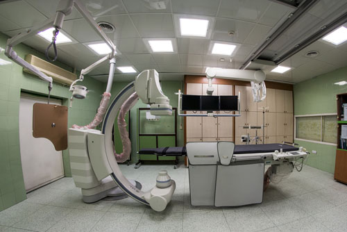
Procedure
Angiograms are always carried out in the catheterization lab of a hospital. Before the procedure starts, your health care team reviews your medical history and asks you about the allergies and medications you take. The team may also do a physical exam and check your vital signs such as blood pressure and pulse.
Conducting angiogram usually takes about an hour. However, it might be longer specially if combined with other cardiac catheterization surgeries. Undoubtedly, preparation and post-procedure care add more time to the whole process. In the very beginning, you are asked to lie on your back on an X-ray table. Since the table might be tilted during the procedure, safety straps will be probably fastened across your chest and legs. X-ray cameras move around your head and chest to take pictures from various angles.
An IV line will be inserted into a vein in your arm. Your doctor may suggest a sedative or some medications and fluids through the IV to help you feel comfortable. You usually become very sleepy but you will be able to be easily awakened to follow the instructions. Electrodes on your chest can closely monitor your heart throughout the process. A blood pressure cuff also tracks your blood pressure and another device, called a pulse oximeter, measures the amount of oxygen in your blood. A small incision is made at the entry site. Then, a catheter is inserted into your artery and carefully threaded to your heart. Afterwards, your physician will inject dye (contrast material) which is easy to see on X-ray images through the catheter. As it moves in the blood vessels, your doctor can observe its flow and identify any blockages or constricted areas. Depending on the discoveries during angiogram, you may need to have more catheter procedures like a balloon angioplasty or a stent placement simultaneously to open up a narrowed artery.
When the procedure is completely performed, your physician removes the catheter from your arm or groin and closes the incision with a clamp or a small plug.
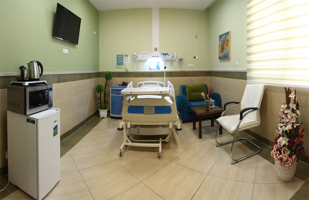
Recovery
After an angiogram, you are usually allowed to go home the same day. Nonetheless, sometimes you are recommended to stay in hospital overnight. You need to drink plenty of fluids to help flush the dye from your body. In case of hunger, you can eat some food only if your doctor approves it. Your health care team will inform you of the time when you can continue taking certain medications, showering, working, and doing other daily activities. It’s better to ask someone to take care of you for a day because the more you rest, the better you will feel. Keep in mind that doing strenuous exercises and heavy lifting are forbidden for at least several days. Your puncture site will possibly remain tender for some days. It might be a bit bruised and have a small bump. There is no need to worry about it since the swelling fades away after a while.
The Results
Doctors can use an angiogram for various reasons. Some of the main ones are mentioned below:
- It shows doctors what's wrong with your blood vessels and how many of your coronary arteries are blocked or narrowed by fatty plaques
- It identifies where blockages are located in your blood vessels precisely
- It demonstrates how much blood flow is blocked through your blood vessels
- It checks the results of previous coronary bypass surgery and the blood flow through your heart and blood vessels
Knowing this information is highly beneficial for doctors since it helps them determine what kind of treatment is the most suitable one for tackling your heart-related problems. It also shows how much danger your heart condition poses to your health. Considering the angiography results, your doctor might decide, for instance, that you need to have coronary angioplasty to clear clogged arteries. To achieve favorable results, doctors may prefer conducting angioplasty or stenting during the procedure to avoid needing another angiogram.
بالنسبة لبعض الإجراءات مثل تصوير الأوعية الدموية ، يجب أن تبقى بدون طعام لمدة أربع ساعات قبل العملية ، وهذا ليس ضرورياً تماماً حيث يُنصح بشرب السوائل ولكن لا يُسمح عادةً بتناول الطعام قبل الإجراء. غالباً ما يتم إجراء الفحوصات المخبرية اللازمة مثل تحاليل الدم وتحديد وظائف الكلى واختبارات تخثر الدم مسبقاً. لا داعي للقلق بشأن مثل هذه المشكلات لأن طبيبك سيخبرك بجميع الإرشادات التي يجب اتباعها بعناية.
حددت صور الأوعية المقطعية بدقة 85٪ من الأفراد الذين يعانون من تضيق كبير و 90٪ من المرضى غير المصابين بمرض الشريان التاجي حتى الآن. يُذكر أن تصوير الأوعية المقطعي المحوسب غير الباضع يكاد يكون دقيقاً مثل تصوير الأوعية التقليدي.
يمكن أن يُظهر تصوير الأوعية الدموية بسهولة ما إذا كانت الشرايين التاجية متضيقة وتحديد موقع تضيقها ومقدار ذلك. يمكن أن يساعد طبيبك أيضاً في معرفة ما إذا كان التغيير في العلاج مثل الأدوية ، أو رأب الأوعية ، أو جراحة مجازة الشريان التاجي مفيداً لتحسين الذبحة الصدرية لديك أو تقليل مخاطر الإصابة بنوبة قلبية أو الوفاة بسبب مشاكل متعلقة بالقلب.
إلى جانب تصوير الأوعية الدموية ، هناك العديد من الاختبارات لفحص الشرايين بما في ذلك:
• فحص الكوليسترول
• الأشعة السينية الصدر
• الإشعة المقطعية
• الموجات فوق الصوتية
• مخطط تخطيط القلب أو اختبار إجهاد القلب
• تخطيط القلب الكهربي
• التصوير بالرنين المغناطيسي أو التصوير المقطعي بالإصدار البوزيتروني
كل من هذه الإجراءات الطبية مرتبطة بالأوعية الدموية. أثناء استخدام تصوير الأوعية الدموية لفحص أو فحص الأوعية الدموية الخاصة بك بحثاً عن مشكلة قلبية محتملة ، يتم استخدام قسطرة لتوسيع الشرايين الضيقة لعلاج الحالة.
سيتحدث معك طبيبك أو جراحك بالتأكيد حول هذه القضايا. في الغالب ، يُسمح بمياه الشرب حتى ساعتين قبل الإجراء.
تسمى العملية التي يتم فيها فحص الأوعية الدموية الشريانية بحثاً عن انسداد محتمل في الدورة الدموية تصوير الأوعية الدموية. تسمى الصور أو القراءات الناتجة عن هذا الإجراء بالصورة الوعائية.
من المحتمل أن يتم علاج الشرايين التاجية الضيقة أثناء تصوير الأوعية الدموية بطريقة تُعرف باسم رأب الوعاء. يتم إدخال قسطرة خاصة عبر الأوعية الدموية إلى الشرايين التاجية للتخلص من الانسداد. البديل الجراحي الآخر للشرايين التاجية الضيقة بشدة هو إجراء المجازة.
يعتبر تصوير الأوعية بشكل عام عملية آمنة ، ولكن الآثار الجانبية البسيطة معتادة وهناك خطر ضئيل بحدوث مضاعفات خطيرة. ستخضع للعملية فقط إذا كانت الفوائد تفوق أي مخاطر محتملة. تحدث إلى جراحك للتعرف على مخاطر إجراء تصوير الأوعية.
معظم الأفراد لا يعانون من مشاكل أثناء أو بعد تصوير الأوعية الدموية.
نادراً ما تحدث النوبات القلبية والسكتات الدماغية ، لكنها قد تكون مهددة للحياة. من المحتمل أن تحدث عند كبار السن أو الأشخاص الذين يعانون من ظروف صحية تزيد من خطر الإصابة بسكتة قلبية أو نوبة قلبية.
تشمل المشكلات الأخرى التي يمكن أن تحدث أثناء تصوير الأوعية الدموية أو بعدها قريبًا ما يلي:
- انسداد مفاجئ أو تمزق في أحد الشرايين.
- تلف الكلى المتعلق بالصبغة.
- كدمات أو نزيف في مكان وضع القسطرة.
- رد فعل تحسسي متعلق بالصبغة المستخدمة لفحص الشرايين.